
CBP® GOALS OF CARE
CBP® Technique emphasizes optimal posture and spinal alignment as the primary goals of chiropractic care while simultaneously documenting improvements in pain and functional based outcomes. The uniqueness of CBP® treatment is in structural rehabilitation of the spine and posture. In general the goals of CBP® Care are:
Normal Front & Side View Posture
Center of mass of head, rib cage & pelvis vertically aligned in Front and Side views.
Normal Spinal Alignment
Front view: vertical alignment
Side View: Harrison Ideal or Average Spinal Model
Normal function
Improved Range of Motion and quality of movement,
Improved muscle strength,
Improved Health & Symptom Improvements
Neck disability index
Oswestry low back index,
SF 36 or Health Status Questionnaire
IDEAL POSTURAL ALIGNMENT:
Ideal postural alignment is depicted in both the frontal and side views. In each view, the center of mass of the skull, thorax, and pelvis are in a vertical line with respect to gravity. In the frontal view, the spinal column is vertically aligned-a straight column- with respect to gravity. In the side view, the spine has three primary curvatures which will be described below:
Neck Curve – Cervical Lordosis
Ribcage Curve – Thoracic Kyphosis
Low back Curve – Lumbar Lordosis
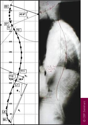
IDEAL SPINAL ALIGNMENT: HARRISON FULL SPINE MODEL
As in all fields of study dealing with the human body, i.e. physiology, hematology, anatomy, etc., there exist normal values for alignment of the spine. The Harrison Spinal Model is an evidenced based model for side view spinal alignment. It is the geometric path of the posterior longitudinal ligament or the backs of the vertebra from the 1st neck vertebra to the bottom of the lower back or top of the sacrum.
CBP® researchers have extensively published ideal and average models for the human spinal curvatures as viewed from the side. This research has lead to the finding of the ‘Harrison Spinal Model’. This model details both Ideal and Average geometric shapes for the curves of the spine from the side. Additionally, ideal and average ranges for the spinal segmental angles for each of the spinal regions have been identified. The neck or cervical spine should have a geometric shape that approximates a ‘piece of a circle’. The ribcage or thoracic spine should have a geometric shape that approximates an oval-elliptical shape. And the low back or lumbar spine should have a geometric shape that approximates an oval-elliptical shape.
These are “evidence based” models. In fact, the CBP® neck-cervical circular model and the low back- lumbar elliptical model have both been found to have discriminative validity between pain and non pain subjects. In other words, the Harrison Spinal Model has been found to be able identify pain subjects versus non-pain subjects by what their spinal x-ray shapes are.
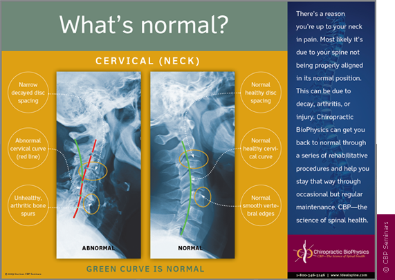
The Harrison Spinal Model in the Neck-Cervical region. The Harrison spinal model is depicted as the GREEN curved line in this figure. On the Right is a normal curved patient x-ray. On the Left is an abnormal curved patient x-ray; where the patient’s abnormal shape is shown by the Red dashed line. The Harrison Spinal Model in the neck has been shown to reasonably predict which person will have neck pain compared to normal subjects.
X-RAY ANALYSIS AND UTILIZATION
To establish optimal and average sagittal models, X-ray analysis and line drawing procedures are utilized. CBP® protocols require that the doctor must measure the displacements on spinal radiographs (segmental Subluxation). Both lateral-side view and anterior to posterior (AP) or frontal view CBP® X-ray line drawing procedures have been studied and found to be reliable. Furthermore, CBP® utilizes standardized x-ray positioning procedures that have been studied and found to be reliable.
As with measures of pain intensity, range of motion, and quality of life, periodic assessment of spinal structural alignment is important to evaluate progress and determine when maximum patient improvement has been reached. In CBP® Technique, the use of initial and follow-up spinal x-rays or radiographs is deemed necessary. Importantly, there is data to show that the use of medical/chiropractic x-rays constitutes a very minor health risk and in fact has been shown to be of benefit (decreased sickness and cancer mortality rates) in some studies.
CBP® POSTURAL ANALYSIS
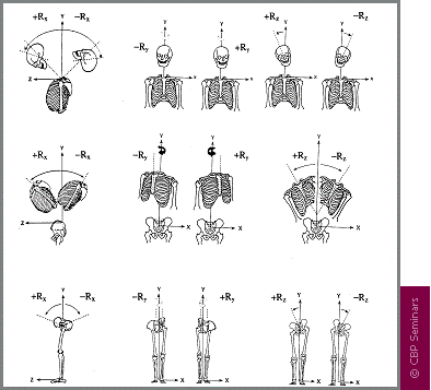
Previously, engineering concepts were used to describe all spinal-vertebral segmental movements as rotations and translations in 3-dimensions. However, Dr. Don Harrison was the first to describe abnormal postures of the head, rib cage, and pelvis in this manner. There are twelve simple motions in six-degrees of freedom as rotations and translations of the human head, ribcage, and pelvis:
• Rotation is a turning, twisting, or tilting movement and is an angular movement.
• Translation is a straight line movement (up, down, left, right, forward, backward).
MIRROR IMAGE® POSTURAL ADJUSTMENTS
In March 1980, Dr. Don Harrison originated postural Chiropractic adjusting procedures that he coined Mirror Image®. Clinically, these adjusting set-ups were found to result in postural and spinal alignment improvements verified with follow up x-ray.
Dr. Don Harrison and his brother Glenn Harrison originated drop table adjustments, instrument adjustments (both table and hand-held). For these new Mirror Image® patient positions, Dr. Don Harrison placed the patient in their opposite posture. These Harrison Mirror Image® positions can be described as reflecting the patient’s head, rib cage, and/or pelvis across the median-sagittal plane in the AP view, and positioning the head, rib cage, and/or pelvis across the mid-frontal plane in the lateral view.
Mirror Image Adjustments, assists the Chiropractor in the rehabilitation of the patient’s posture. In theory, these adjustments re-balance the bodies sense of proper balance or alignment by way of triggering improved muscle and nerve reflexes. Thus, postural adjustments as performed with drop table, hand-held instrument, or even mirror image manipulation procedures, are performed for resetting the nervous system regulation of postural muscle balance.
MIRROR IMAGE® POSTURAL EXERCISES
Further, in 1980-1986, Dr. Don and Dr. Glenn Harrison began developing and testing Mirror Image® posture exercises. Mirror image® exercises are performed to stretch shortened muscles and to strengthen those muscles that have weakened in areas where postural muscles have adapted to asymmetric abnormal postures. Although strength and conditioning exercise has not proven to correct posture, postural exercises performed in the mirror image have shown initial promise in the reduction of posture and spinal displacements.
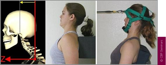
MIRROR IMAGE® POSTURAL AND SPINAL TRACTION
Additionally from 1980-1986, for use in difficult cases, Dr. Don Harrison and Dr. Glenn Harrison originated several Mirror Image® postural traction (rotations and translations of the head, rib cage, and pelvis) and cervical extension (backwards bending) traction methods to restore the sagittal cervical curve. These CBP® Technique cervical extension traction methods were improved upon over the years by several other CBP® practitioners.
From 1996-2000, several postural and spinal traction methods to restore thoracic and lumbar sagittal curvatures were developed by Dr. Deed Harrison; Dr. Don Harrison’s son. Dr. Deed began to further refine the CBP® Technique cervical traction methods with an analysis of head posture, curve configuration, thoracic curvature, gender, and body size. From 1992-2004, six non-randomized clinical control trials, one randomized clinical control trial, and five case studies have been performed on these CBP® Technique Mirror Image® procedures.
Postural mirror image and extension traction for the side view spinal curves provides sustained loading periods of 10-20 minutes and is necessary to cause visco-elastic deformation to the resting length of the spinal ligaments, muscles, and discs. In clinical controlled trials, extension traction, as done in CBP® Technique, is the only proven method (without surgery) shown to consistently correct abnormal spinal curvatures back towards their normal alignment.
There are MANY types of spinal extension traction methods used in CBP® Technique; some are more aggressive and technical than others. Each patient’s spine is unique and thus, a variety of traction devices are used depending upon the exact condition of the patient.
TREATMENT INTERVENTIONS
CBP® structural rehabilitation procedures include Mirror Image® exercises, adjustments, and traction (referred to as the E.A.T protocol). The mirror image® posture positions are the rotation and translation pairs in or about each coordinate axis. Thus, CBP® care is multi-modal and is consistent with Bolton’s ideas of clinical applications in evidence based practice.
The reason for postural mirror image exercises, adjustments, and traction procedures is to address all the tissues involved in spine and posture alignment.
Unlike the pain management care, the E.A.T corrective care protocol is unique to CBP®.
REFERENCES
1 Harrison DD. CBP Technique: The Physics of Spinal Correction. National Library of Medicine #WE 725 4318C, 1982-97. Harrison DD. CBP Technique: The Physiscs of Spinal Correction.
2 Harrison DD. Spinal Biomechanics: A Chiropractic Perspective. National Library of Medicine #WE 725 4318C, 1982-97. Harrison DD. CBP Technique: The Physiscs of Spinal Correction.
3 Harrison DD. CBP X-ray Work Book. National Library of Medicine #WE 725 4318C, 1982-97. Harrison DD. CBP Technique: The Physiscs of Spinal Correction.
4 Harrison DE, Harrison, DD, Haas JW. CBP® Structural Rehabilitation of the Cervical Spine. Evanston, WY: Harrison CBP Seminars, 2002.
5 Harrison DE, Harrison, DD, Haas JW, Oakley PA. Spinal Biomechanics for Clinicians, Volume I. Evanston, WY: Harrison CBP Seminars, 2003.
6 Harrison DE, Harrison, DD, Haas JW, Oakley PA. CBP® Structural Rehabilitation of the Lumbar Spine. Evanston, WY: Harrison CBP Seminars, 2005.
7 Harrison DE, Betz J, Harrison, DD. CBP® Structural Rehabilitation of the Thoracic Spine. Evanston, WY: Harrison CBP Seminars, 2005.
8 Chiropractic Biophysics Online. http://www.idealspine.com/. Services/research papers. Accessed 12-15-04.
9 Harrison DD, Janik TJ, Harrison GR, Troyanovich SJ, Harrison DE, Harrison SO. Chiropractic Biophysics Technique: A Linear Algebra Approach to Posture in Chiropractic. J Manipulative Physiol Ther 1996;19(8):525-535.
10 Panjabi MM, White AA, Brand RA. A Note on Defining Body Parts Configurations. J Biomech 1974; 7:385.
11 Harrison DE, Harrson DD. The Lumbar Spine: Rehabilitation with the CBPâ method. National Library of Medicine #WE 725 4318C, 1997.
12 Harrison DE, et al. Changes in sagittal lumbar configuration with a new method of extension traction. Presented at the 27th annual meeting of the International Society for the Study of the Lumbar spine. Adelaide, Australia April 9-13, 2000;151.
13 Harrison DE, Harrison DD, Cailliet R, Janik TJ, Holland B. Changes in Sagittal Lumbar Configuration with a New Method of Extension Traction: Non-randomized Clinical Control Trial. Arch Phys Med Rehab 2002;83(11):1585-1591.
14 Harrison DD, Jackson BL, Troyanovich SJ, Robertson GA, DeGeorge D, Barker WF. The Efficacy of Cervical Extension-Compression Traction Combined with Diversified Manipulation and Drop Table Adjustments in the Rehabilitation of Cervical Lordosis. J Manipulative Physiol Ther 1994;17(7):454-464.
15 Harrison DE, Harrison DD, Betz J, Colloca CJ, Janik TJ, Holland B. Increasing the Cervical Lordosis with Seated Combined Extension-Compression and Transverse Load Cervical Traction with Cervical Manipulation: Non-randomized Clinical Control Trial. J Manipulative Physiol Ther 2003; 26(3): 139-151.
16 Harrison DE, Cailliet R, Harrison DD, Janik TJ, Holland B. New 3-Point Bending Traction Method of Restoring Cervical Lordosis Combined with Cervical Manipulation: Non-randomized Clinical Control Trial. Arch Phys Med Rehab 2002; 83(4): 447-453.
17 Harrison DE, Cailliet R, Betz JW, Harrison DD, Haas JW, Janik TJ, Holland B. Harrison Mirror Image Methods for Correcting Trunk List: A Non-randomized Clinical Control Trial. Eur Spine J 2004; In Press.
18 Harrison DE, Harrison DD, Haas JW, Betz JW, Janik TJ, Holland B. Conservative Methods to Correct Lateral Translations of the Head: A Non-randomized Clinical Control Trial. J Rehab Res Devel 2004;41(4):631-640.
19 Zaleski BW, Wood J. A comparison of Palmer package and Chiropractic Biophysics for the treatment of pain. Proceedings of the International Conference on Spinal Manipulation 1992; Chicago, IL., May 15-17.
20 Paulk GP, Bennett DL, Harrison DE. Management of a chronic lumbar disk herniation with CBP methods following failed chiropractic manipulative intervention. J Manipulative Physiol Ther 2005;in press.
21 Ferrantelli JR, Harrison DE, Harrison DD, Steward D. Conservative management of previously unresponsive whiplash associated disorders with CBP methods: a case report. J Manipulative Physiol Ther 2005;in press.
22 Haas JW, Harrison DE, Harrison DD, Bymers B. Reduction of symptoms in a patient with syringomyelia, cluster headaches, and cervical kyphosis. J Manipulative Physiol Ther 2005;in press.
23 Harrison DE, Bula J. Non-operative correction of the flexible flat back using lumbar extension traction: A case study of three. J Chiropractic Education 2002;16(1).
24 Bastecki A, Harrison DE, Haas JW. ADHD: A CBP case study. J Manipulative Physiol Ther 2004; 27(8):e14.
25 Harrison DE, Harrison DD. Chiropractic Biophysics Technique. Part I: The Scientific Basis. Eur Chiropractic J 2005;
26 Harrison DD, Janik TJ, Troyanovich SJ, Holland B. Comparisons of Lordotic Cervical Spine Curvatures to a Theoretical Ideal Model of the Static Sagittal Cervical Spine. Spine 1996;21(6):667-675.
27 Harrison DD, Harrison DE, Janik TJ, Cailliet R, Haas JW, Ferrantelli J, Holland B. Modeling of the Sagittal Cervical Spine as a Method to Discriminate Hypo-Lordosis: Results of Elliptical and Circular Modeling in 72 Asymptomatic Subjects, 52 Acute Neck Pain Subjects, and 70 Chronic Neck Pain Subjects. Spine 2004; 29:2485-2492.
28 Janik TJ, Harrison DD, Cailliet R, Troyanovich SJ, Harrison DE. Can the Sagittal Lumbar Curvature be Closely Approximated by an Ellipse? J Orthop Res 1998; 16(6):766-70.
29 Harrison DD, Cailliet R, Janik TJ, Troyanovich SJ, Harrison DE, Holland B. Elliptical Modeling of the Sagittal Lumbar Lordosis and Segmental Rotation Angles as a Method to Discriminate Between Normal and Low Back Pain Subjects. J Spinal Disord 1998; 11(5): 430-439.
30 Harrison DE, Janik TJ, Harrison DD, Cailliet R, Harmon S. Can the Thoracic Kyphosis be Modeled with a Simple Geometric Shape? The Results of Circular and Elliptical Modeling in 80 Asymptomatic Subjects. J Spinal Disord Tech 2002; 15(3): 213-220.
31 Harrison DD, Harrison DE, Janik TJ, Cailliet R, Haas JW. Do Alterations in Vertebral and Disc Dimensions Affect an Elliptical Model of the Thoracic Kyphosis? Spine 2003; 28(5):463-469.
32 Harrison DE, Harrison DD, Cailliet R, Troyanovich SJ, Janik TJ. Cobb Method or Harrison Posterior Tangent Method: Which is Better for Lateral Cervical Analysis? Spine 2000; 25: 2072-78.
33 Harrison DE, Cailliet R, Harrison DD, Janik TJ, Holland B. Centroid, Cobb or Harrison Posterior Tangents: Which to Choose for Analysis of Thoracic Kyphosis? Spine 2001; 26(11): E227-E234.
34 Harrison DE, Harrison DD, Janik TJ, Harrison SO, Holland B. Determination of Lumbar Lordosis: Cobb Method, Centroidal Method, TRALL or Harrison Posterior Tangents? Spine 2001; 26(11): E236-E242.
35 Harrison DE, Holland B, Harrison DD, Janik TJ. Further Reliability Analysis of the Harrison Radiographic Line Drawing Methods: Crossed ICCs for Lateral Posterior Tangents and AP Modified Risser-Ferguson. J Manipulative Physiol Ther 2002; 25: 93-98.
36 Harrison DE, Harrison DD, Colloca CJ, Betz J, Janik TJ, Holland B. Repeatability of Posture Overtime, X-ray Positioning, and X-ray Line Drawing: An Analysis of Six Control Groups. J Manipulative Physiol Ther 2003; 26(2): 87-98.
37 Haas M, Taylor JAM, Gillette RG. The routine use of radiographic spinal displacement analysis: a dissent. J Manipulative Physiol Ther 1999;22:254-259.
38 Phillips RB. Plain film radiography in chiropractic. J Manipulative Physiol Ther 1992;15:47-50.
39 Taylor JAM. Full-spine radiography: a review. J Manipulative Physiol Ther 1993;16:460-474.
40 Kauffman JM. Diagnostic radiation: are the risks exaggerated? J Amer Phys Surg 2003;8(2):54-55.
41 Cohen BL, Lee IS. A catalog of risks. Health Physics 1979;36:707-722.
42 International Atomic Energy Agency. Facts about low-level radiation. American Nuclear Society, 1982.
43 Janik TJ, Harrison DE, Cailliet R, Harrison DD, Normand MC, Black P. Validity of an algorithm to estimate 3-D rotations and translations of the head in upright posture from three 2-D digital images. 2005; In Review.
44 Harrison DE, Janik TJ, Cailliet R, Harrison DD, Normand MC, Black P, Perron DL. Validation of an algorithm to estimate 3-D rotations and translations of the thorax and pelvis in upright posture from three 2-D digital images. 2005 In Review.
45 Bolton JE. The evidence in evidence-based practice: what counts and what doesn’t count? J Manipulative Physiol Ther 2001;24:362-366.
46 Hrysomallis C, Goodman C. A review of resistance exercise and posture realignment. J Strength Cond Res 2001;15:385-390
47 Den Boer WA, Anderson PG, Limbek JV, Kooijman MAP. Treatment of idiopathic scoliosis with side shift therapy: An initial comparison with a brace treatment historical cohort. European Spine J 1999;8:406-410.
48 Weiss HR. Influence of an in-patient exercise program on scoliotic curve. Ital J Orthop Traumatol 1992; 18:395-406.
49 Weiss HR, Lohschmidt K, el-Obeidi N, Verres C. Preliminary results and worst-case analysis of in patient scoliosis rehabilitation. Pediatr Rehabil 1997;1:35-40.
50 Pearson ND, Walmsley RP. Trial into the effects of repeated neck retractions in normal subjects. Spine 1995; 20(11): 1245-51.).
51 DeVocht JW, Pickar JG, Wilder DG. EMG activity levels of paraspinal muscles during spinal manipulation. Proceedings from the 29th annual meeting of ISSLS 2002; Vancouver, Canada.
52 Oliver MJ, Twomey LT. Extension creep in the lumbar spine. Clin Biomech 1995;10:363-368.
53 Harrison DE, Harrison DD, Troyanovich SJ. Three-Dimensional Spinal Coupling Mechanics. Part II: Implications for Chiropractic Theories and Practice. J Manipulative Physiol Ther 1998; 21(3): 177-86.
54 Harrison DE, Cailliet R, Harrison DD, Troyanovich SJ, Janik TJ. Cervical Coupling on AP Radiographs During Lateral Translations of the Head Creates an S-Configuration. Clin Biomech 2000; 15(6): 436-440.
55 Harrison DE, Cailliet R, Harrison DD, Janik TJ, Troyanovich SJ, Coleman RR. Lumbar Coupling During Lateral Traction.